Contents
01
A Harsh Reality — Complications Are the Wake-Up Call
Complications including vascular embolism, tissue necrosis and visual impairment are widely reported.
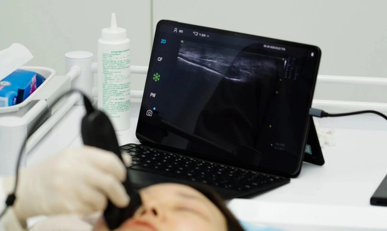
Portable ultrasound may well become the third eye for aesthetic physicians.
02
Far More Complex Than Many Realize — Four High-Risk Facial Zones
Facial vascular networks exhibit remarkable anatomical complexity.
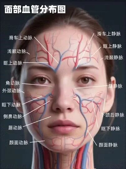
- Glabella / Forehead: Supratrochlear and supraorbital arteries — risk of irreversible blindness.
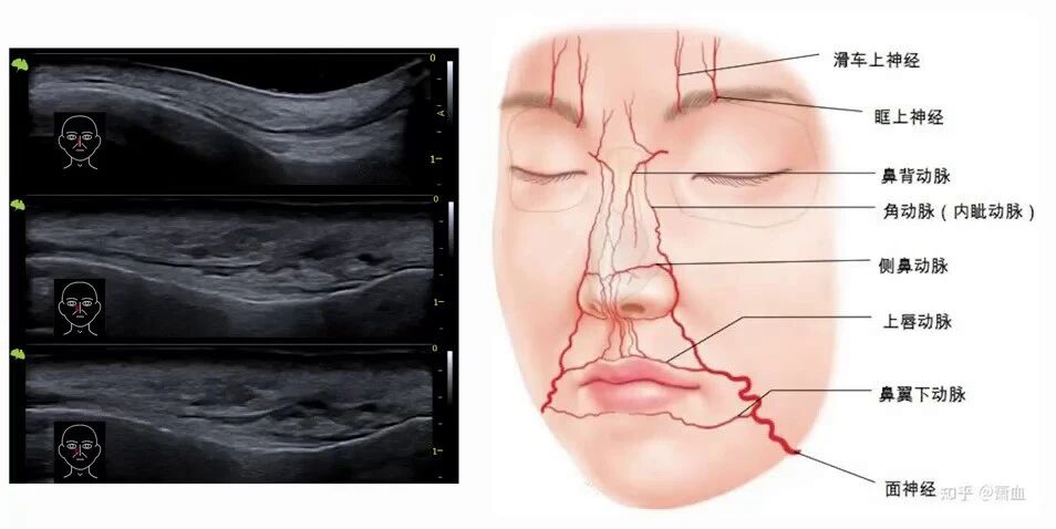
- Dorsum Nasi / Nasal Tip: Dorsal nasal artery — identical risks.
- Cheek / Malar Region: Superficial facial and zygomaticofacial arteries — easy needle penetration.
- Lips: Variable superior and inferior labial arteries — frequent embolism sites.
These vessels range merely 0.5–2 mm in diameter — visualization is fundamental.
03
The Cost of Over-Filling — Origins & Treatment Challenges
If vascular embolism is the acute crisis, "pillow-face deformity" is the chronic challenge.

Origins of Pillow-Face Deformity
- Over-filling: Excessive volume per session or repeated high-frequency injections.
- Incorrect injection planes: Fillers deposited in superficial fat compartments.
- Inappropriate product selection.
- Aesthetic misjudgment.
- Vicious cycle of revision.
Challenges in Treating Pillow-Face Deformity
- Hyaluronidase dissolution.
- Surgical curettage and aspiration.
- Regenerative agents (PLLA etc.).
- Expectant management.
Prevention outweighs treatment — prevention depends on visualization.
04
Physicians' Third Eye — A Clinical Look from Daily Practice
A young aesthetic physician shares real-world insights integrating portable ultrasound.
Pre-injection Assessment — Three High-Risk Zones
Glabella, temporal fossa, and nose are the highest-risk regions.
- Glabella: supratrochlear and supraorbital arteries.
- Temporal fossa: up to ten distinct tissue planes.
- Nose: dorsal nasal artery — clinically well-documented embolism.
Ultrasound visualizes vessel locations.
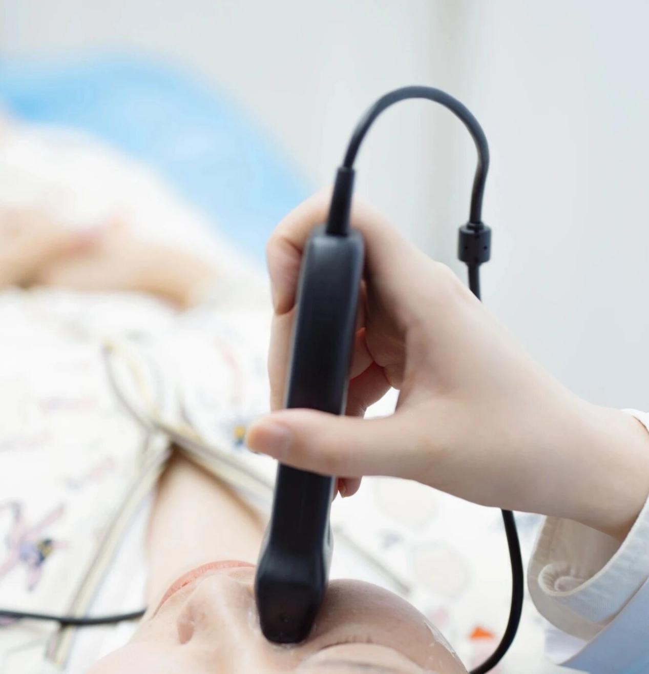
Pillow-Face Revision — Ultrasound Identifies "Invisible" Fillers
Patients frequently cannot specify which injectables they previously received.
- Differentiate filler types via characteristic ultrasound signatures.
- Confirm anatomical planes.
Clinical example: ultrasound revealed a tiny hyaluronic-acid collection beneath the tear-trough ligament.
Full-Workflow Support
- Pre-procedure: distinguish volume-loss vs ptosis-driven ageing.
- Intra-procedure: real-time guidance to target planes.
- Long-term follow-up: assess fat-pad repositioning, fascial tightening.
05
To Inject Precisely, One Must First See Clearly
Aesthetic injections extend far beyond manual craft.
Miniaturization and intelligence are democratizing visualized injection.
Equipping more clinicians with visualization tools is the professional pursuit.