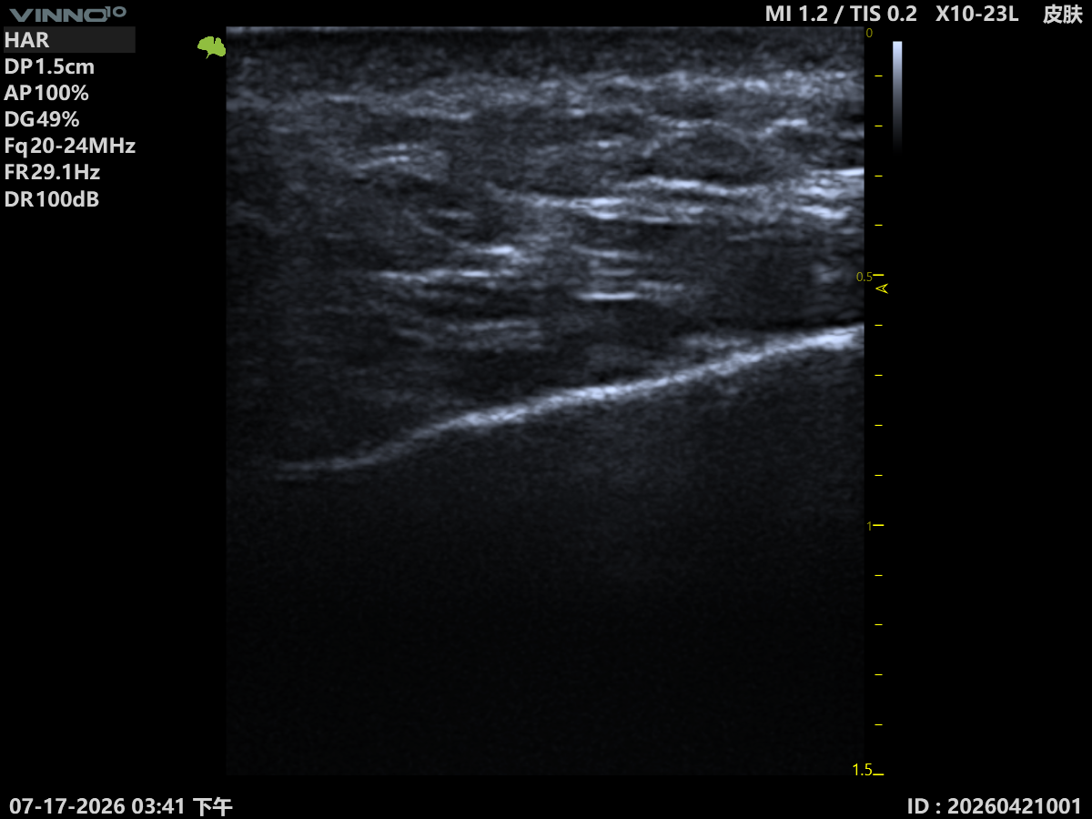
Full-Cycle Visualization · Across the Aesthetic Journey

From pre-procedure assessment to post-treatment management — imaging evidence for every treatment.

Visualizing Every Step of Aesthetic Care

Ultrasound Guidance Across the Treatment Cycle

Pre-op assessment, intra-op guidance, post-op management — imaging support at every stage.
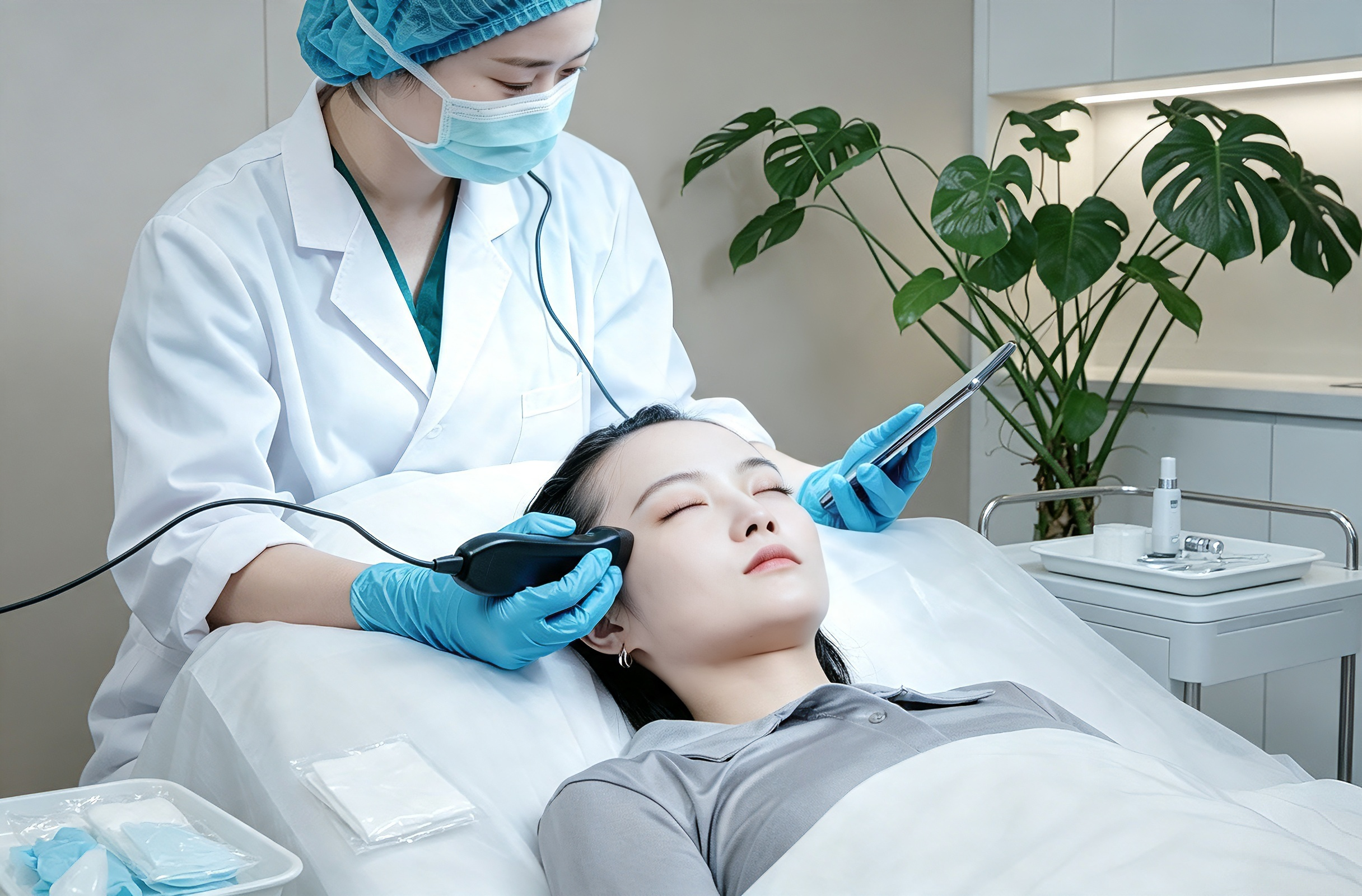
Pre-Procedure Assessment
01

Pre-Procedure Assessment

See the anatomy, plan with precision

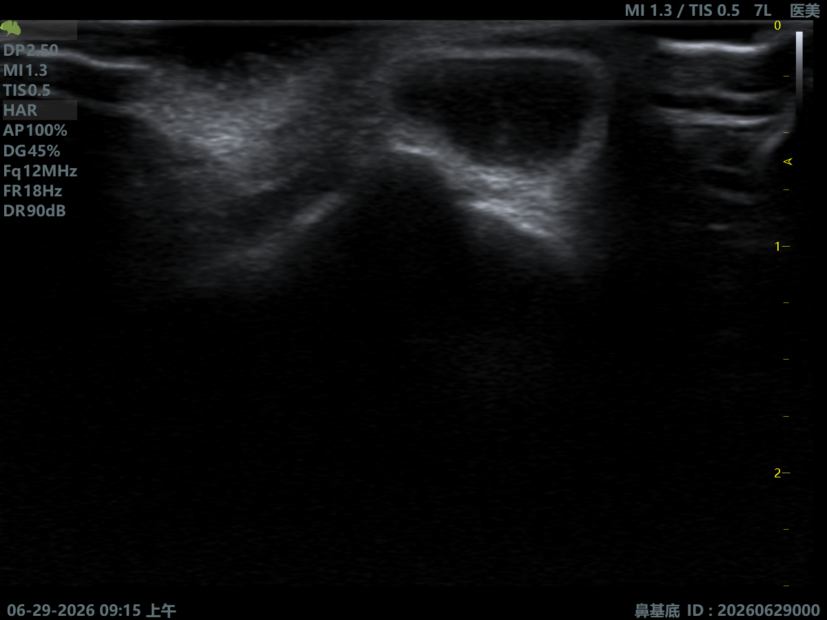
Every patient's vasculature, fat pads and tissue structure differ. High-frequency ultrasound helps physicians understand individual anatomical features before treatment — a reliable basis for injection, anti-aging and minimally-invasive therapy.
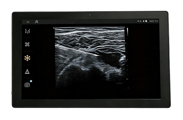
Vascular Mapping Fat-Pad Analysis SMAS Fascia Assessment Facial Aging Analysis Treatment Planning
Injection Therapy
02

Injection Therapy

Real-time imaging for precise injection

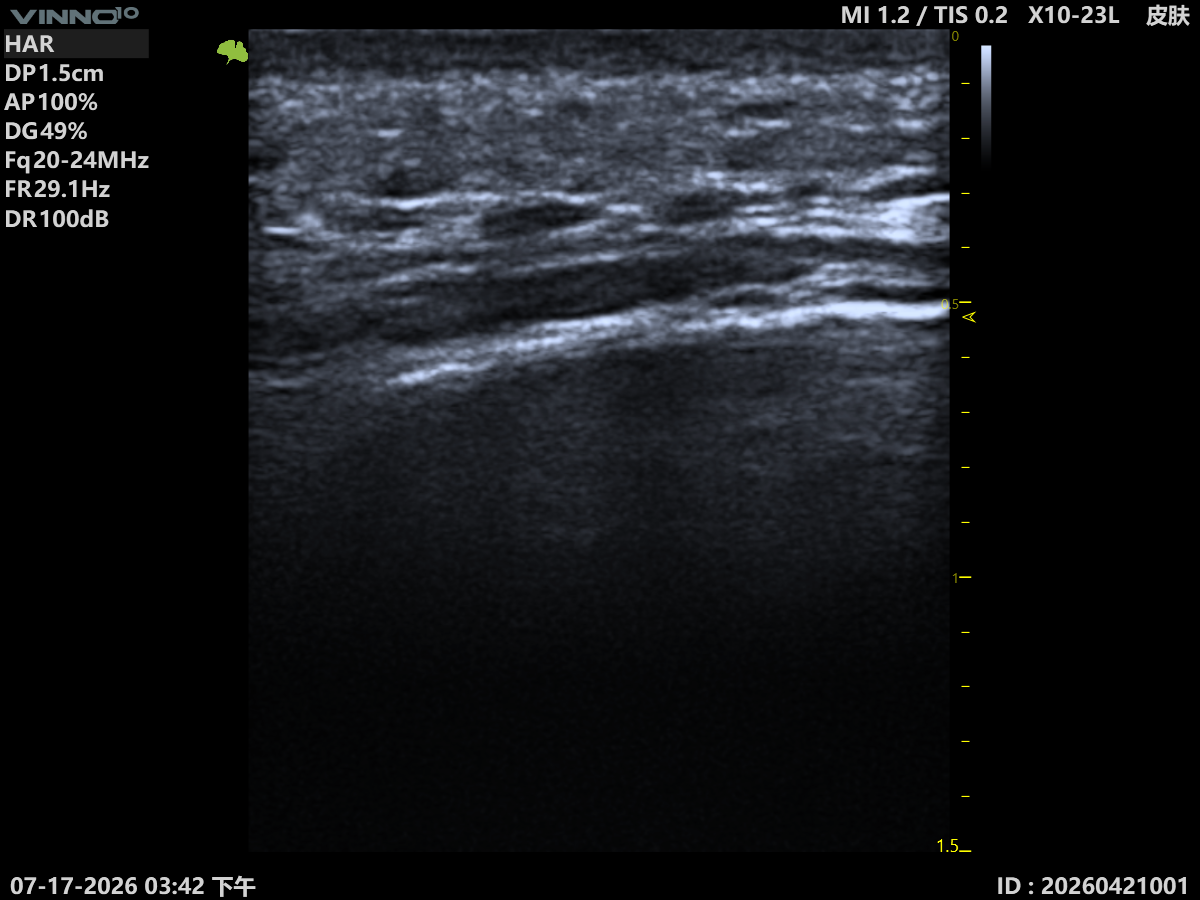
Observe tissue layers and target areas in real time during treatment, helping physicians complete filler, regenerative and other injection procedures with greater precision.
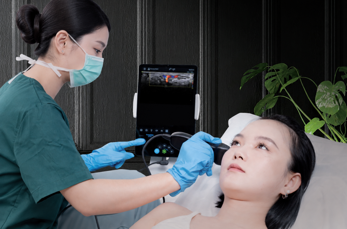
Hyaluronic Acid Filler Regenerative Treatment Botulinum Toxin Mesotherapy & Superficial Injection Real-Time Ultrasound Guidance
Facial Rejuvenation
03

Facial Rejuvenation

Make aging visible

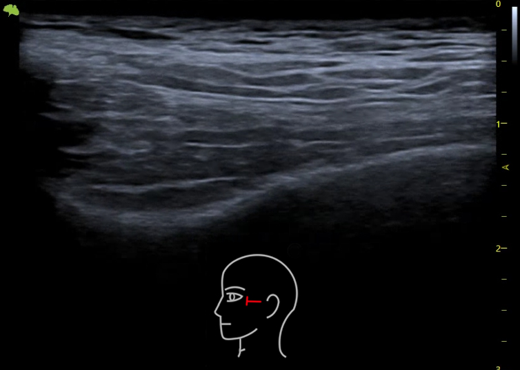
Aging happens beyond the skin surface — fat loss, fascia laxity and deep-tissue change all play a part. Ultrasound helps physicians objectively assess tissue state at every layer, giving rejuvenation therapy a more scientific foundation.
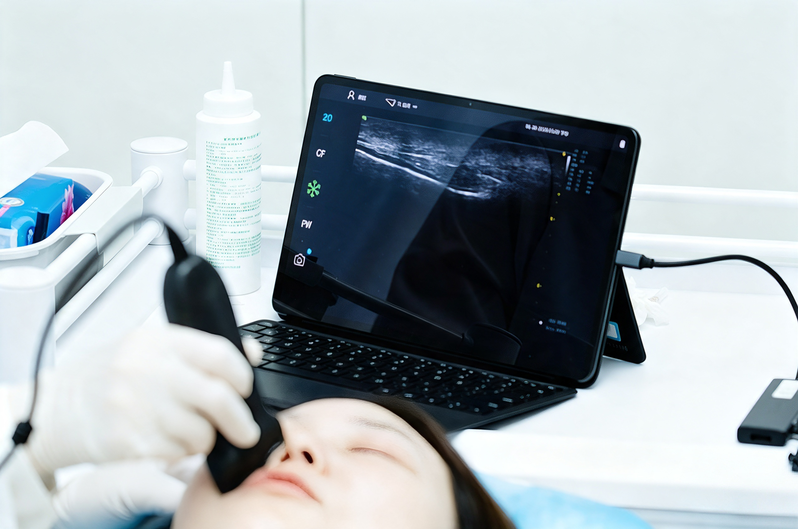
Volume Assessment Tissue Support Assessment Personalized Anti-Aging Plan Outcome Assessment & Follow-up Minimally-Invasive Rejuvenation
Complication Management
04

Complication Management

Detect issues, resolve them more scientifically

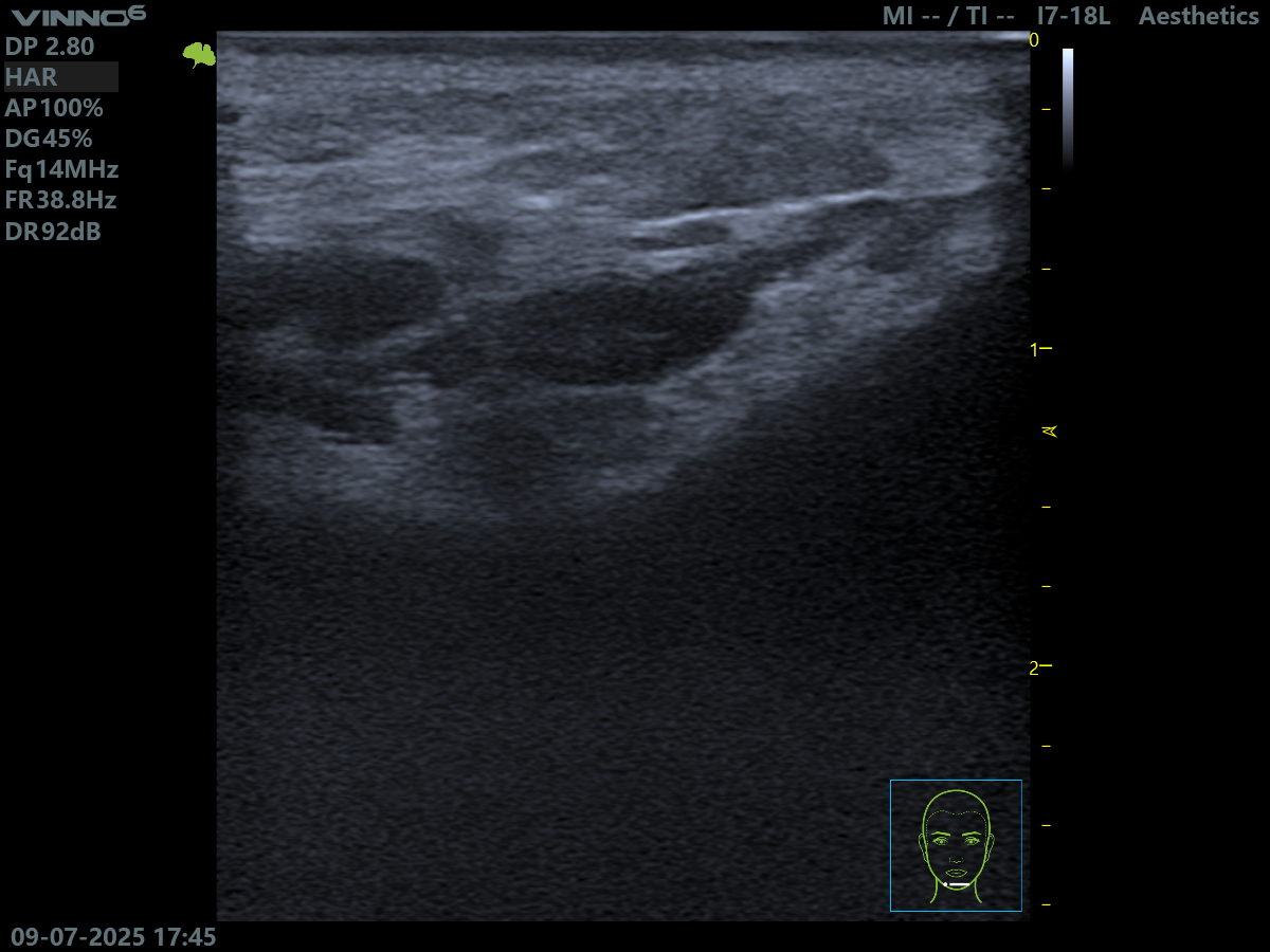
Ultrasound helps physicians objectively assess post-treatment issues and provides reliable evidence for correction — from over-fill to vascular embolism, ultrasound-assisted complication care is more precise.
Over-Fill Syndrome Filler Migration Granuloma & Nodules Inflammation Assessment Ultrasound-Assisted Correction

Pre-Procedure Assessment

Vascular Mapping
Identify key vessel paths and anatomical variants to aid risk assessment and treatment planning.
Fat-Pad Analysis
Observe fat-pad state and volume change as a reference for rejuvenation therapy.
SMAS Fascia Assessment
Analyze facial support structures and tissue state to aid anti-aging design.
Facial Aging Analysis
Assess aging features at the structural level to support personalized decisions.
Treatment Planning
Build a more precise, targeted treatment strategy from imaging assessment.
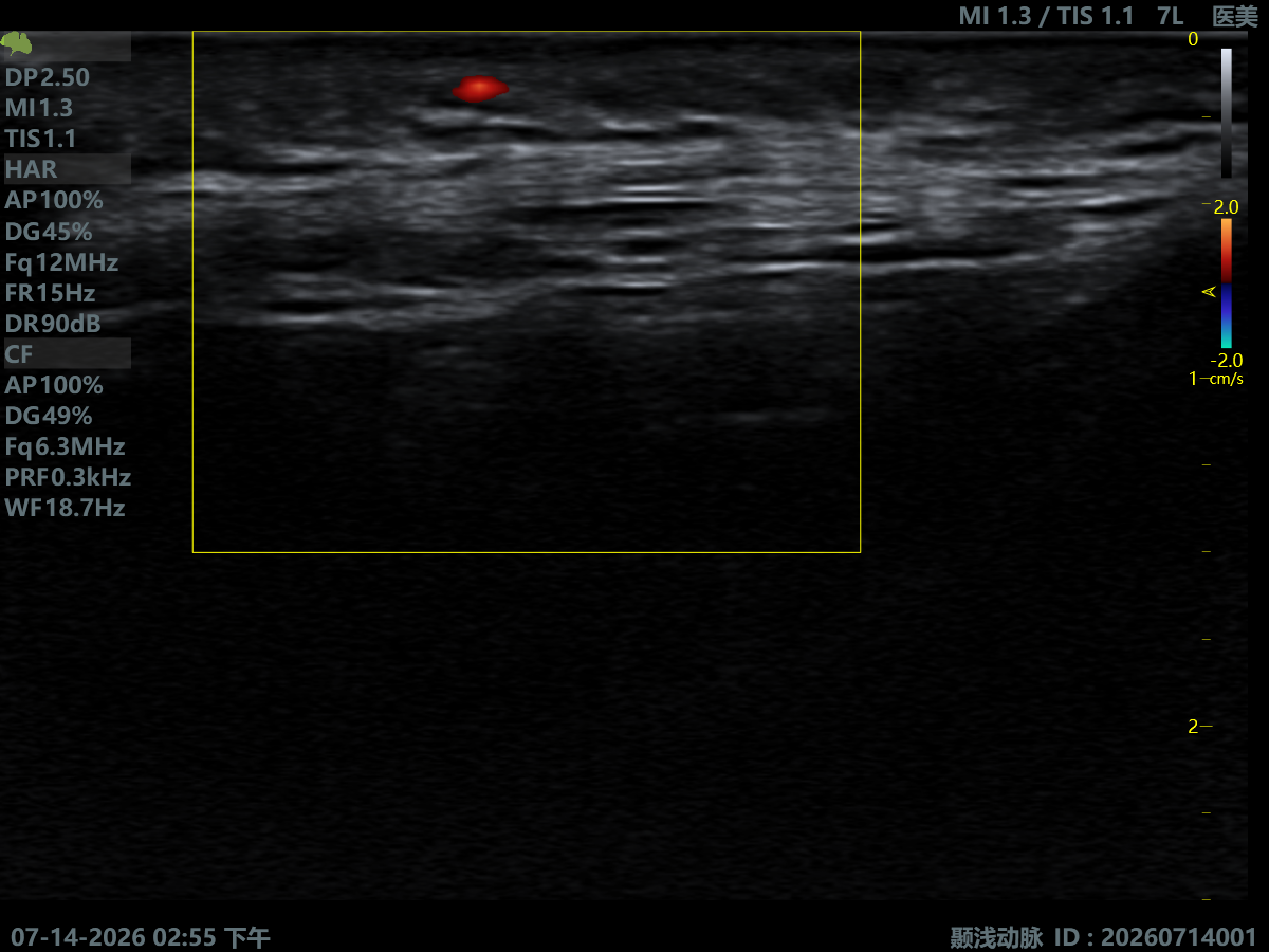
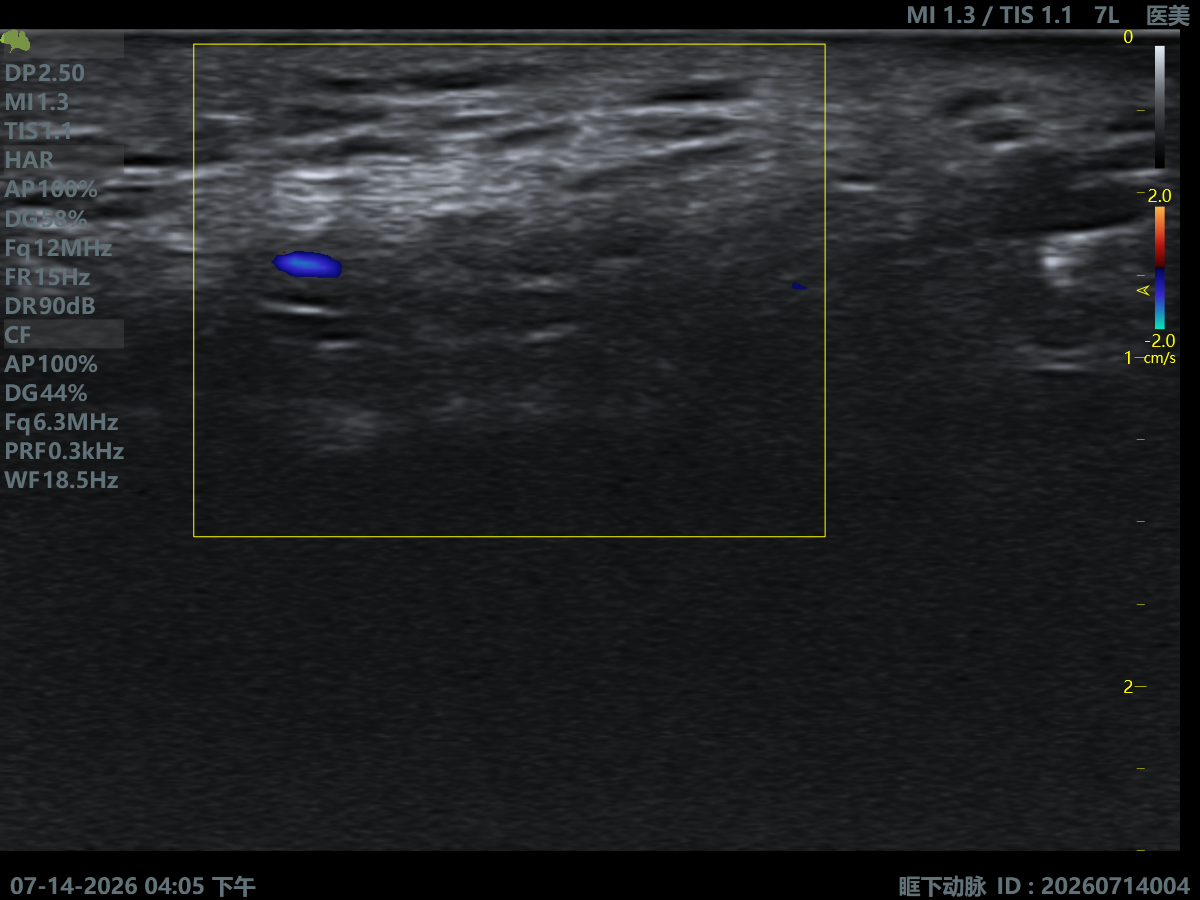
Superficial Temporal Artery
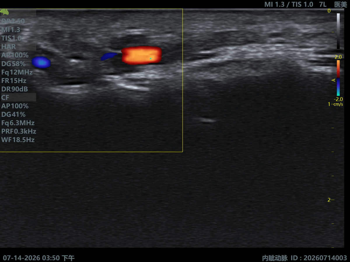
Infraorbital Artery
Infraorbital Artery
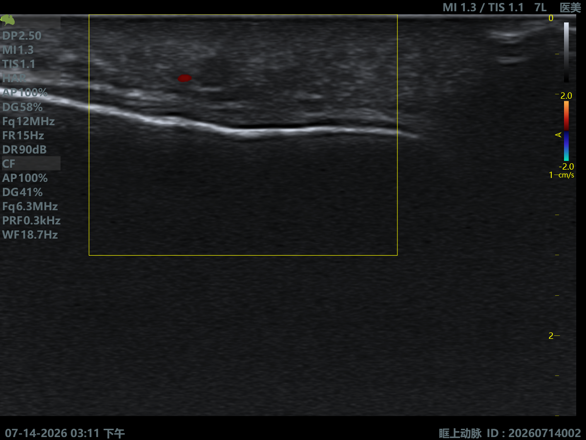
Supraorbital Artery
Supraorbital Artery
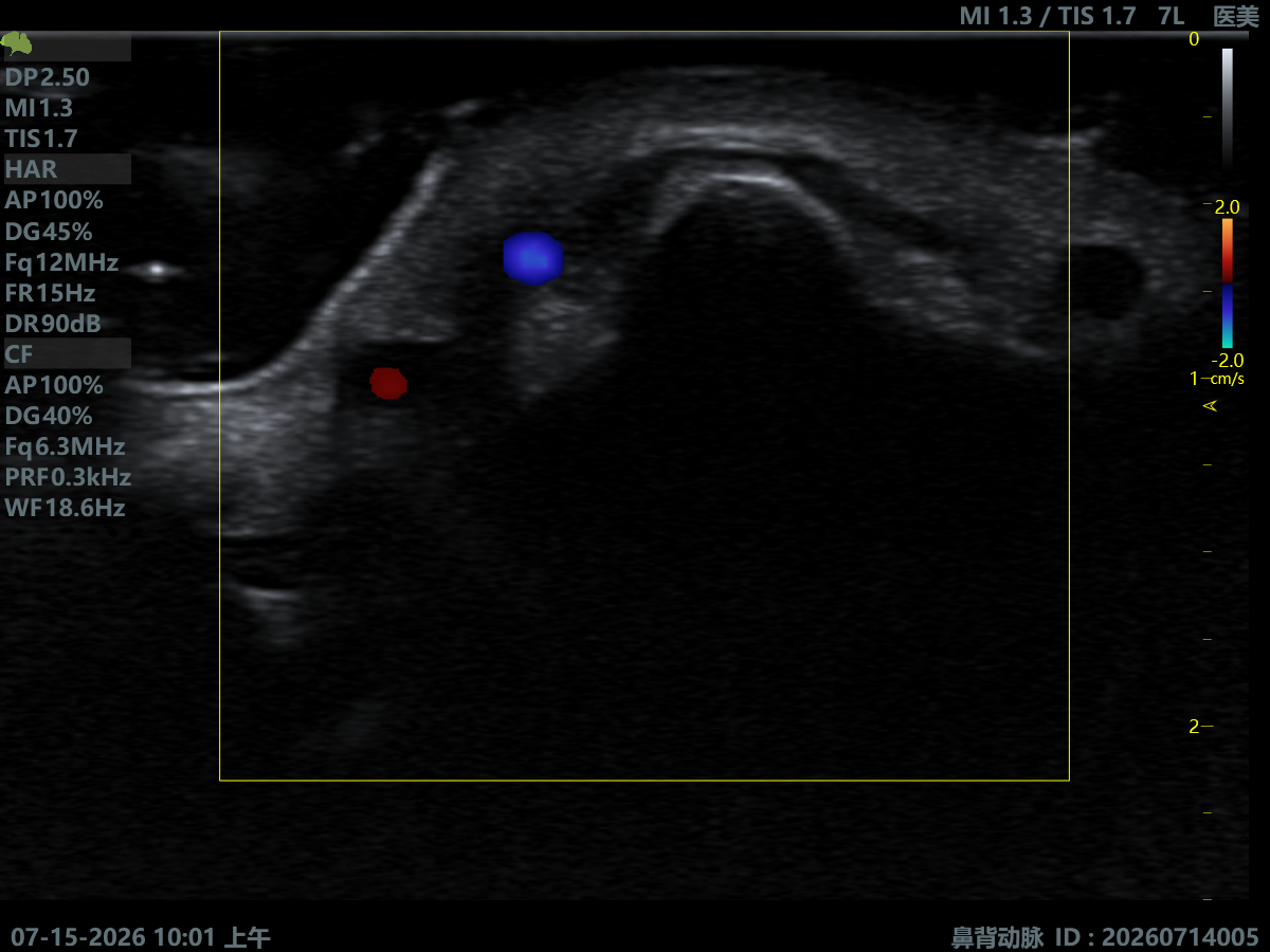
Dorsal Nasal Artery
Dorsal Nasal Artery
Angular Artery & Vein

Facial Rejuvenation

Volume Assessment
Observe fat-pad state and facial volume change to quantify aging.
Tissue Support Assessment
Assess the state of SMAS, ligaments and soft-tissue support structures.
Personalized Anti-Aging Plan
Tailor treatment strategy to anatomical features for precise anti-aging.
Outcome Assessment & Follow-up
Build objective imaging records for long-term tracking and outcome comparison.
Minimally-Invasive Rejuvenation
Aids thread-lift, regenerative and combined-therapy treatment design.
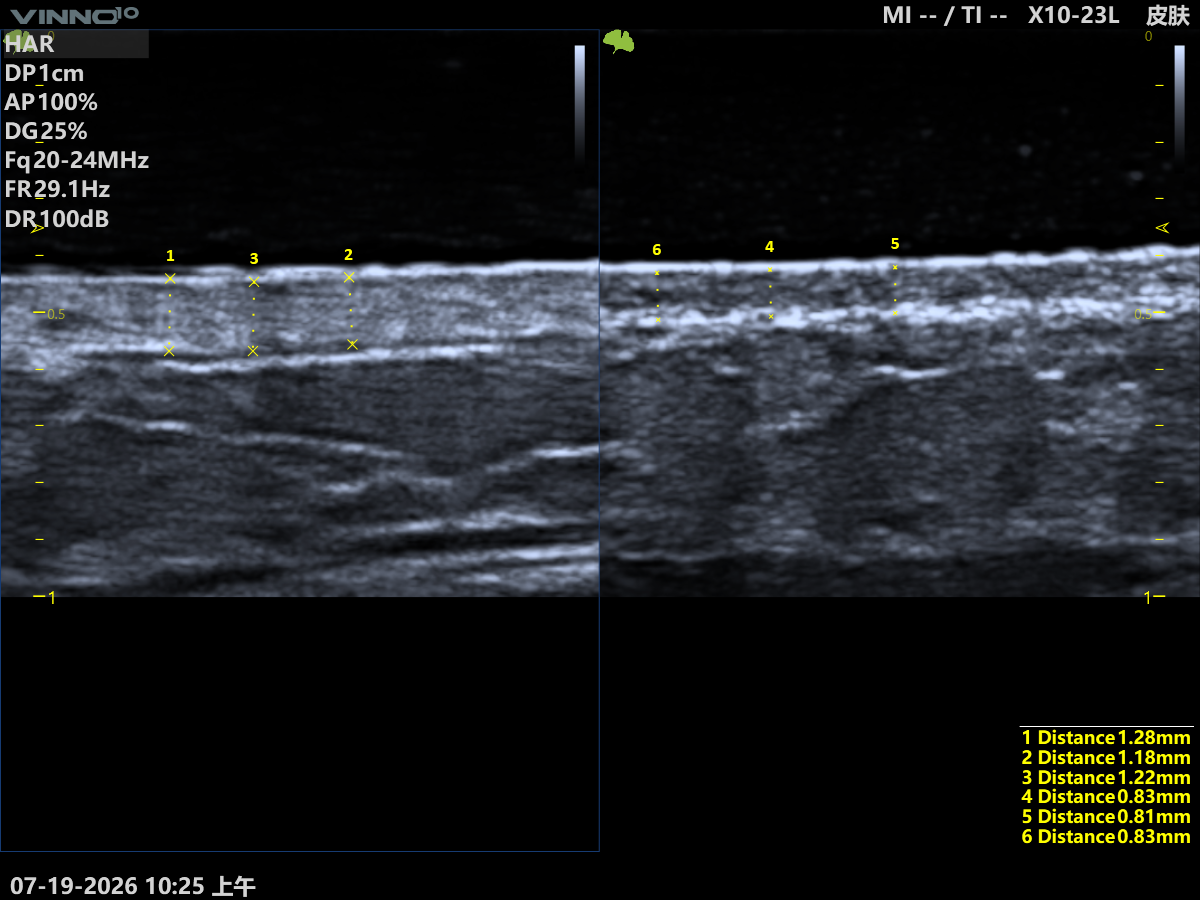
Facial Ultrasound Imaging
Facial Ultrasound Imaging 2

Complication Management

Over-Fill Syndrome
Assess filler accumulation, tissue bulging and contour change.
Filler Migration
Observe filler position and diffusion path to assess migration extent.
Granuloma & Nodules
Identify abnormal tissue reaction and lesion extent to aid diagnosis and treatment.
Inflammation Assessment
Observe tissue inflammation and repair state to objectively assess post-treatment response.
Ultrasound-Assisted Correction
Aids filler localization, treatment planning and correction-process assessment.

Why Aesthetic Ultrasound?

From experience to visualized decision-making

Facial vasculature, fat and soft-tissue structure vary markedly between individuals, so experience alone cannot fully reveal true anatomy. Ultrasound lets physicians see key structures before treatment, get real-time feedback during treatment, and objectively assess afterward — making the whole process more precise, controllable and traceable.
Severe vascular-complication rate reduced
Injection precision improved, filler dosage more controllable
Patient trust and satisfaction markedly improved
  • Improves treatment precision and operator confidence
  • Supports personalized treatment planning
  • Strengthens patient communication and trust
  • Aids post-treatment outcome assessment and management
  • Builds a standardized, visualized clinical workflow
Why Aesthetic Ultrasound
Explore Clinical Applications

Explore Ultrasound in Aesthetic Clinical Practice

From anatomical understanding to clinical practice — build a full-cycle aesthetic ultrasound knowledge system.